
Point-of-care imaging across specialties
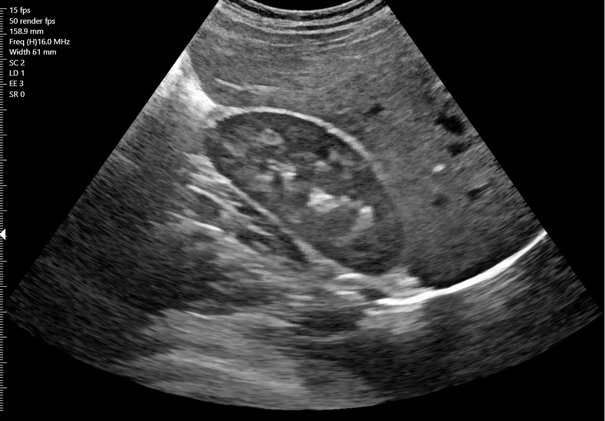
The UltraVision is a high-resolution point-of-care system well suited to visualizing muscles, tendons, ligaments, nerves, vessels and glands throughout the body. Because of its unique design, new applications arrive as software algorithm upgrades.
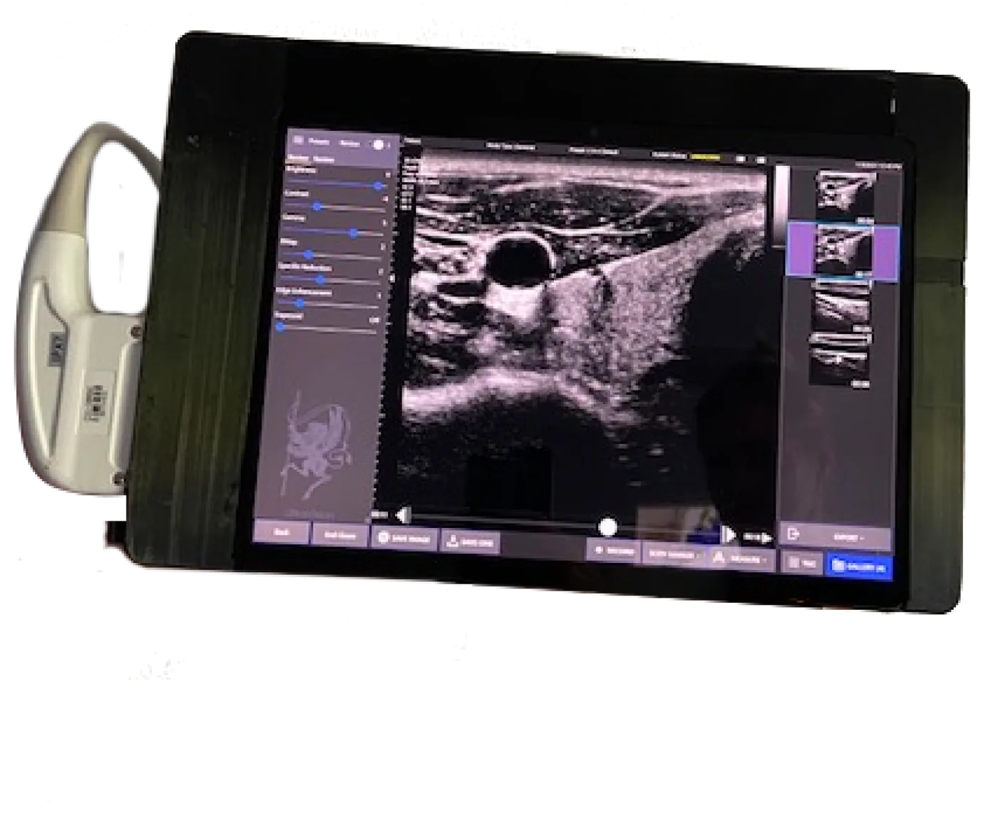
Clinical use now
Within the cleared indications for small parts, superficial musculoskeletal, abdominal and peripheral vessel imaging.
Software upgrades to come
- Obstetrics and gynecology
- Prostate scanning
Future applications are simple software algorithm upgrades to the system you already own.
MSK ultrasound
Musculoskeletal ultrasound is a dynamic, non-invasive exam that allows high-resolution, real-time evaluation of musculoskeletal disorders. It is a cost-effective, reliable modality to assess and diagnose soft tissue conditions, strains, sprains, muscle tears and other disorders, and it is valuable when range of motion is lost through acute or chronic injury to major tendons, in pain management and in urgent care.
The UltraVision's combination of image quality, workflow and features makes it a preferred choice for MSK work: it helps visualize sprains, strains, tears, trapped nerves and arthritis. Needle visualization for guided injections is in clinical development.
Used in orthopedics, rheumatology, physical medicine and rehabilitation, regenerative medicine (precision injection of platelet-rich plasma or stem cells), podiatry, physical therapy, anesthesiology, pain management and sports medicine.
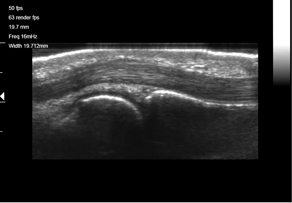
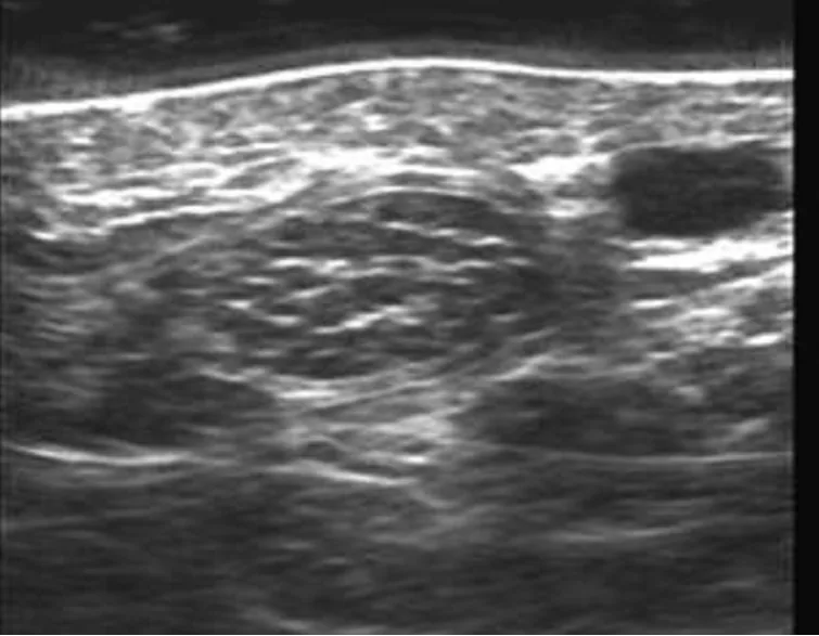
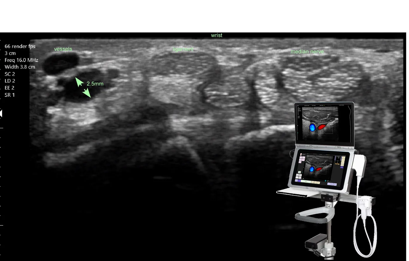
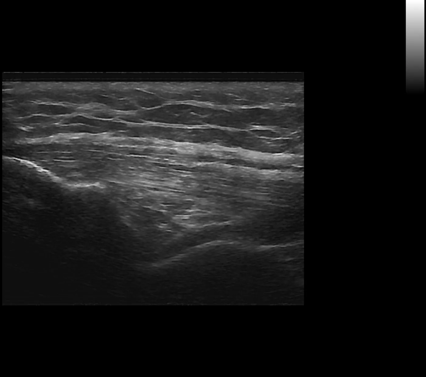
Scanning protocol videos
Short clips recorded on the UltraVision with sonographer narration, by joint. Press play on any clip; nothing downloads until you do.
Learn MSK ultrasound
Mobile learning, in-office training by MSK experts, workshops and the CPT codes that apply to MSK exams and guided injections.
Education and reimbursement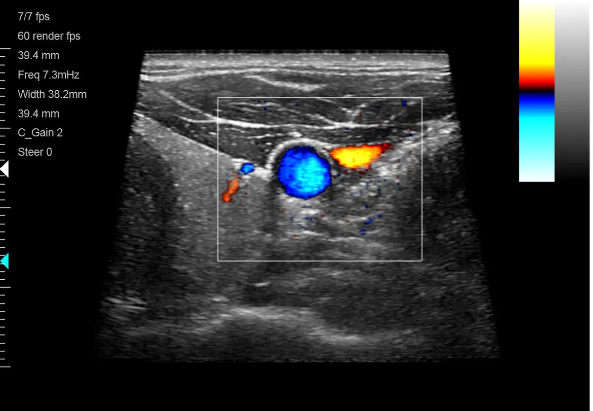
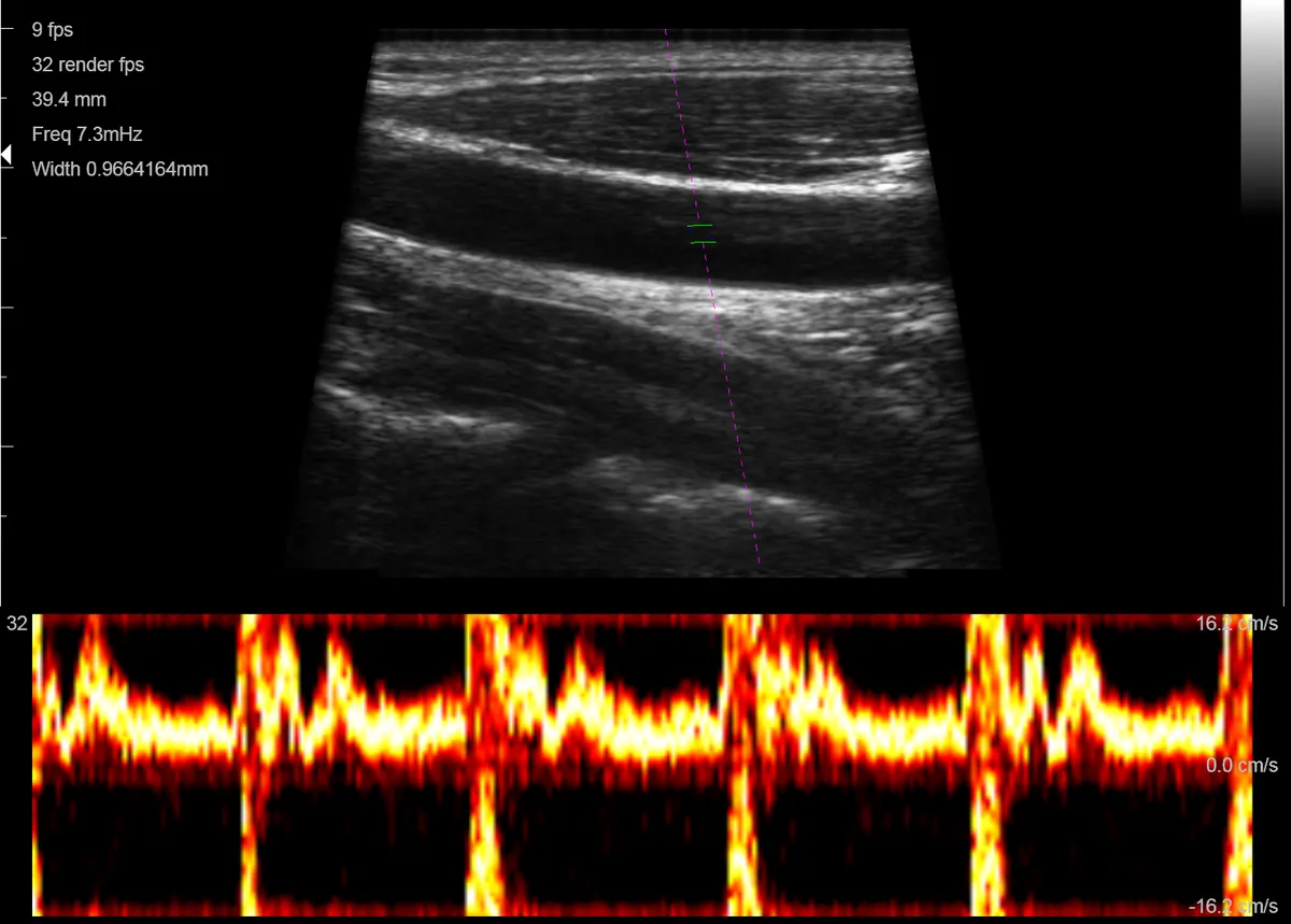
Peripheral vascular imaging
The resolution of the L14-4 linear transducer with colour flow Doppler and pulse-wave Doppler makes the UltraVision ideal for studying the peripheral vascular system. Peripheral veins under 4 mm across are clearly resolved in cross section and longitudinally.
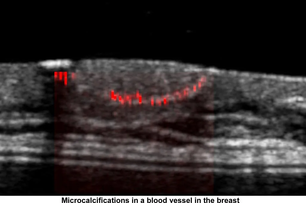
Carotid stenosis and calcified plaque are also visible with our MC-mode research work. Read about MC-mode.
Thyroid and small parts
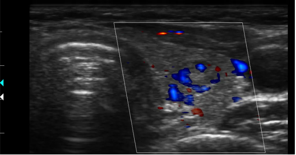
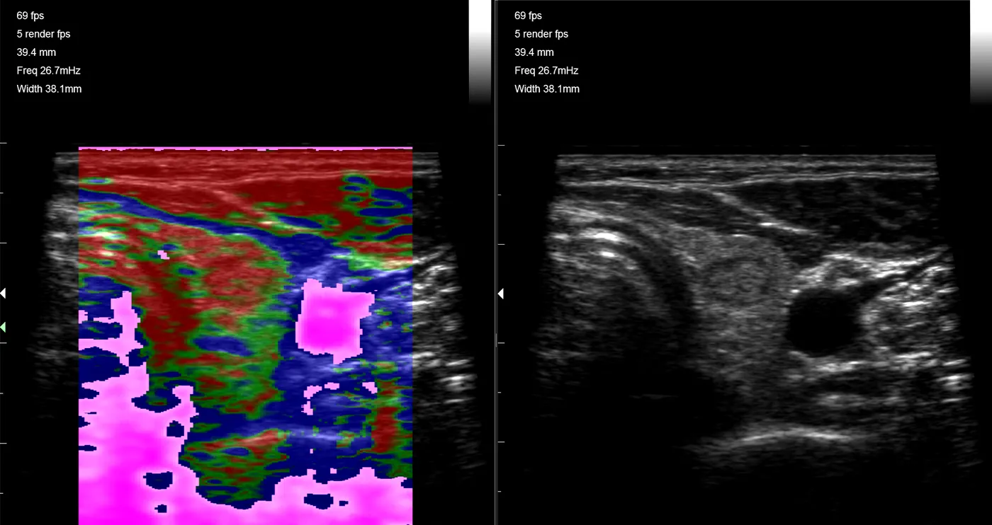
Point-of-care ultrasound lets endocrinologists visualize the thyroid and locate masses: checking for thyroid nodules, identifying fluid-filled cysts and looking for an enlarged or inflamed gland.
The UltraVision uses a proprietary algorithm combining spatial compounding, speckle reduction, reverberation reduction and edge enhancement to increase small-structure resolution and border delineation. Elastography shows the relative stiffness of a nodule. Needle visualization for biopsy and aspiration is in clinical development.
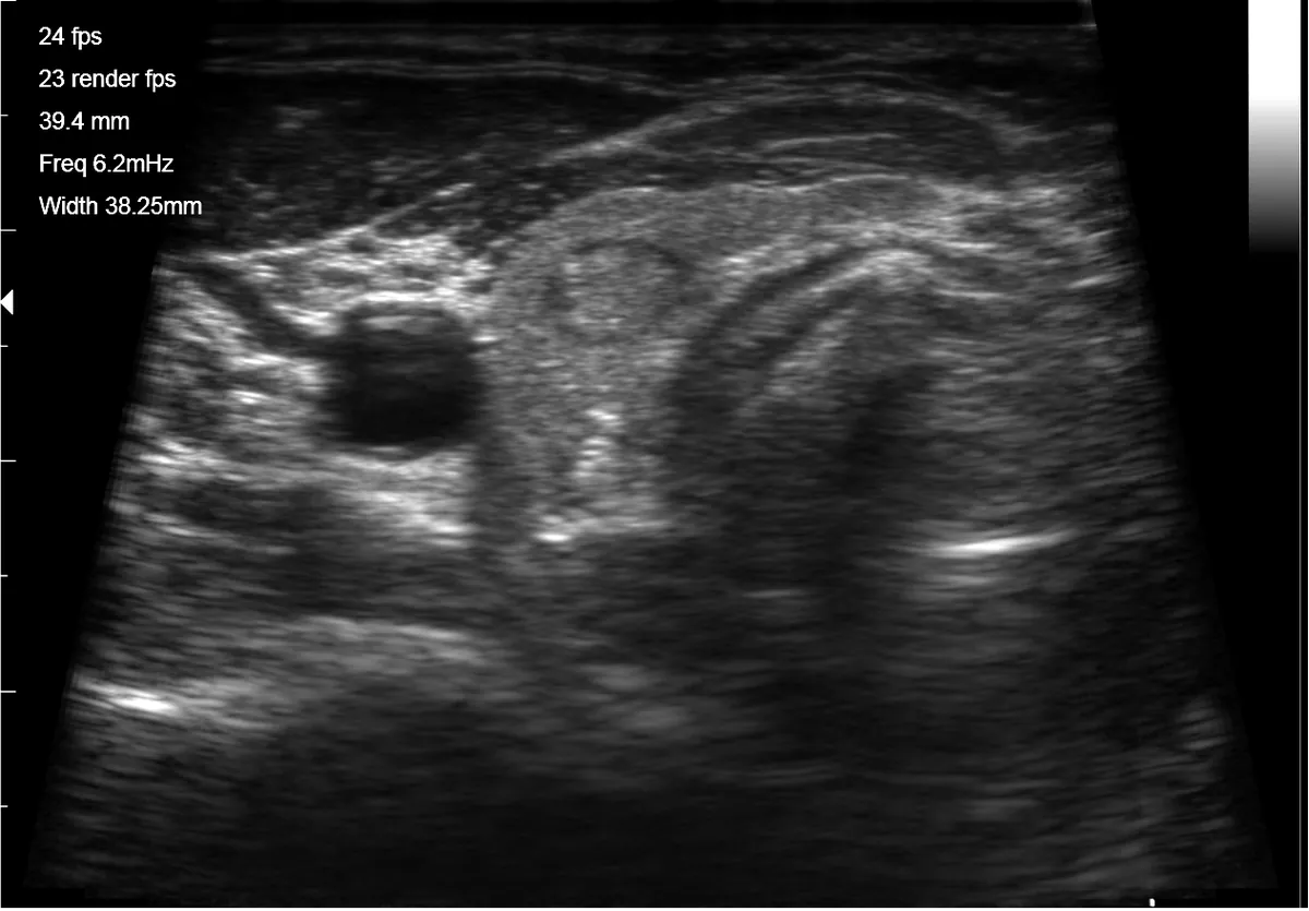
Lower extremity venous imaging
The UltraVision system, paired with the L14-4 linear transducer and simultaneous colour flow Doppler and B-mode imaging, provides high-resolution anatomical visualization and hemodynamic evaluation of peripheral vessels, including lower extremity venous anatomy. We are seeking partners to commercialize phlebology applications.
This application is currently in clinical development and limited by United States law to investigational use (21 CFR 812).
Partner with usNeedle visualization
The user indicates on the touch screen the approximate direction of the needle's approach. The UltraVision then sends specific acoustic lines in that direction, optimized for edge enhancement of the needle's hard acoustic interface, and uses features of the elastographic algorithm to show tissue moving in response to the needle. Ultra-fine needle placement with the surrounding image intact.
This feature is currently in clinical development and limited by United States law to investigational use (21 CFR 812).
On the UltraVision XSPreparing for the breast market
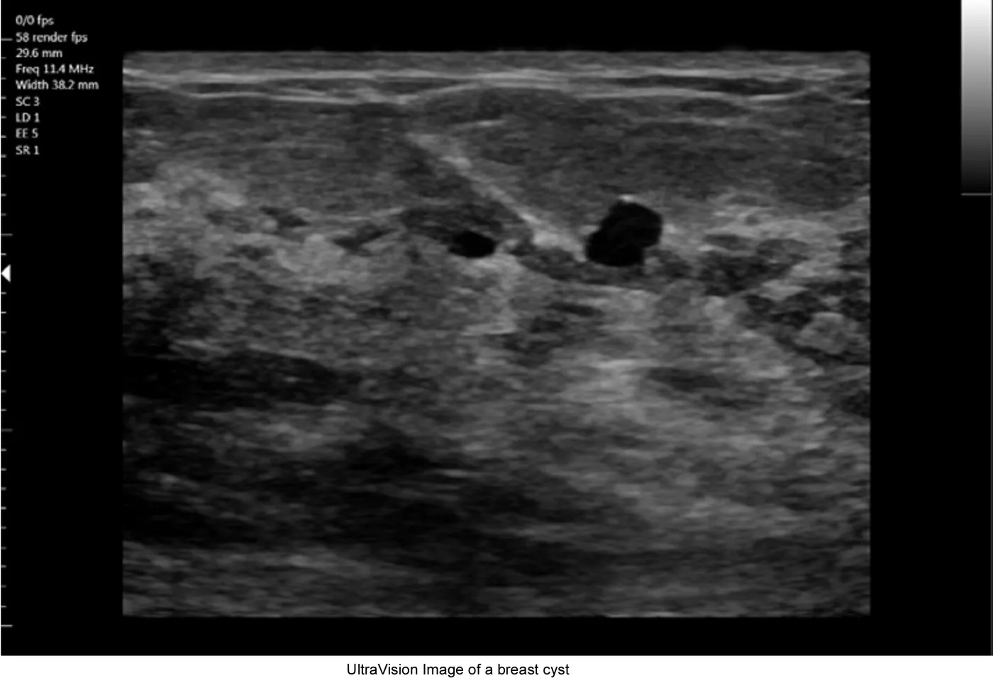
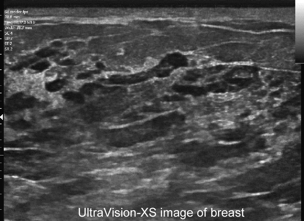
UltraVision is being prepared for breast imaging. The components for this market stand as follows:
- B-mode for solid versus cyst determination: FDA 510(k) cleared and available today
- Fine-needle biopsy guidance: a software algorithm, in clinical development (investigational)
- Colour flow Doppler for evaluating vascularity: cleared and available today
- Compression elastography for relative tissue hardness: cleared and available today
- Shear elastography for quantitative tissue hardness: developed
- Visualization of microcalcifications (MC-mode): U.S. Patent 12,773,182 (October 2026), does not affect acoustic output, investigational and available for research today
- Automatic breast screening attachment: concept phase
We are seeking partners to assist in the development of breast market products. Read about MC-mode.
Bring UltraVision to your specialty
We seek experts in each of the ultrasound specialties to participate with us in bringing point-of-care systems to market.