
UltraVision XS
Designed for the bedside, the private office and the interventional environment. The XS gives the physician enhanced visualization at the point of care for anesthesia, breast, musculoskeletal and urgent care applications.
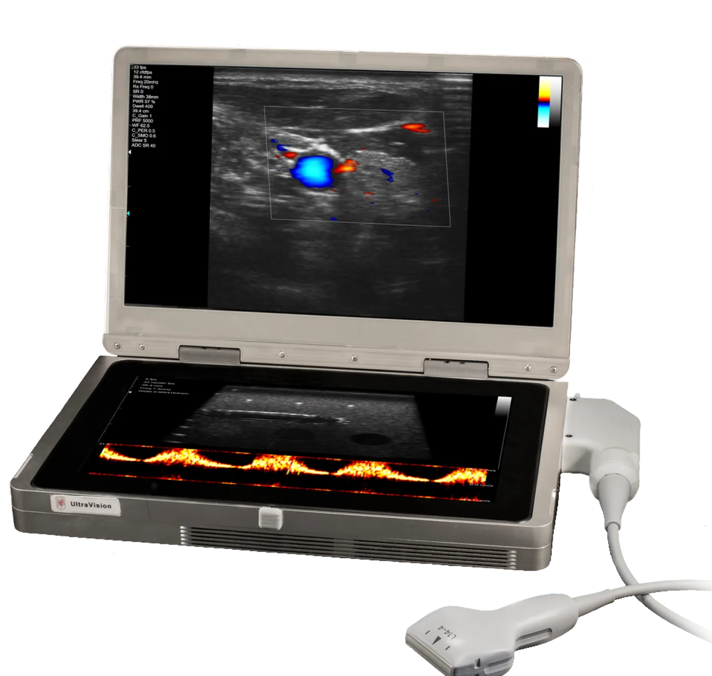
Image quality and workflow, by design
Proprietary algorithms create uncompromised image quality, and practical features shorten procedure time.
UltraBeam compounding
Multiple scan angles create a single image with enhanced contrast resolution. Simultaneous acoustic lines increase the resolution of small structures and sharpen border delineation.
Needle visualization In development
An advanced algorithm sends acoustic lines to intersect the needle and uses elastography to show the tissue it moves, for precise placement without losing the image around it.
This feature is currently in clinical development and limited by United States law to investigational use (21 CFR 812).
Strain elastography
Non-invasive assessment of tissue stiffness in response to transducer pressure. Valuable for assessing change in tendons, ligaments and nodules.
UltraConnect
A Wi-Fi dongle in a USB port connects the system for upgrades, service, support, training and image optimization, wherever it is.
UltraZoom and UltraView
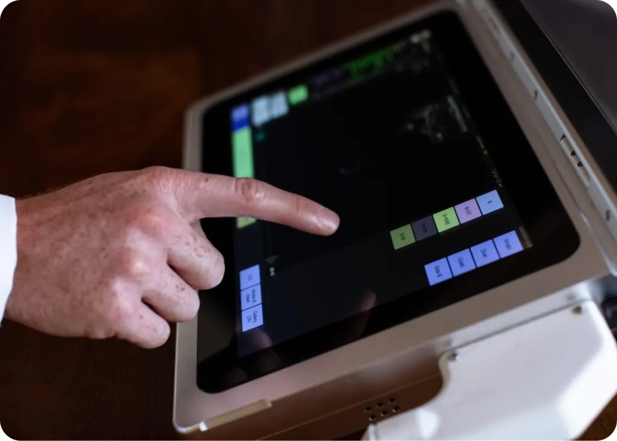
Pinch to zoom down to less than a square centimetre, about 30 times normal. View up to four images at once in duplex or triplex modes.
One-touch optimization
A single key instantly optimizes the image in B, colour and PW modes. Presets programmed by our consultant sonographers deliver excellent images minutes out of the box.
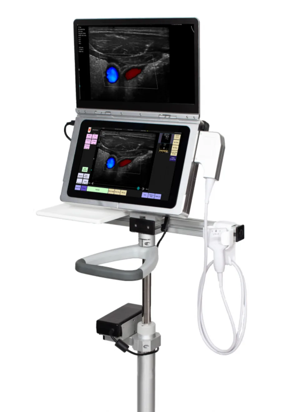
UltraVision XS at a glance
- Viewing display
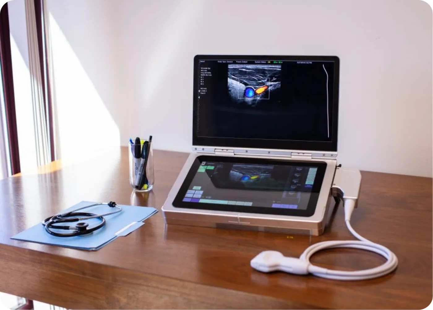
- 15.6″ IGZO, 3000×1800 pixels, paper-white, angle-independent, 1000:1 contrast
- Control display
- 13.3″ HDMI capacitive multi-touch, 1920×1080 pixels, carries all system controls
- Processor
- Intel i7 PC running Windows, 16 GB RAM, 512 GB solid-state drive
- Image processing
- GPU programmed in DirectX plus a single very large FPGA with up to eight simultaneous beamformers
- Transducers
- 2 to 20 MHz linear, curved linear and phased arrays, industry-standard connector
- Modes
- B, M, pulse-wave Doppler, colour flow Doppler, compression elastography, harmonic imaging, photoacoustics. In development: needle visualization
- Storage and export
- SQL patient database with images, cines and recordings; DICOM-compatible output; USB 3 ports; optional external HDMI monitor
- Connectivity
- Any PC keyboard or mouse, Wi-Fi dongle, voice control, remote support
- Power
- IEC 60601-1 desktop supply; optional accessory with up to two user-replaceable batteries, one hour of scanning each, swapped without restarting
- Case and weight
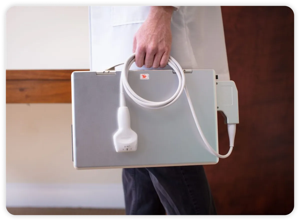
- Solid machined aluminum; 15×10×2 inches closed; 12 lb
- Start-up
- Ready to scan in seconds
- Regulatory
- FDA 510(k) cleared. For prescription use only. Indications for use
- Warranty
- Five years
Specifications are subject to change. Contact us for the current specification sheet and configuration options.
Three processors, one programmable design
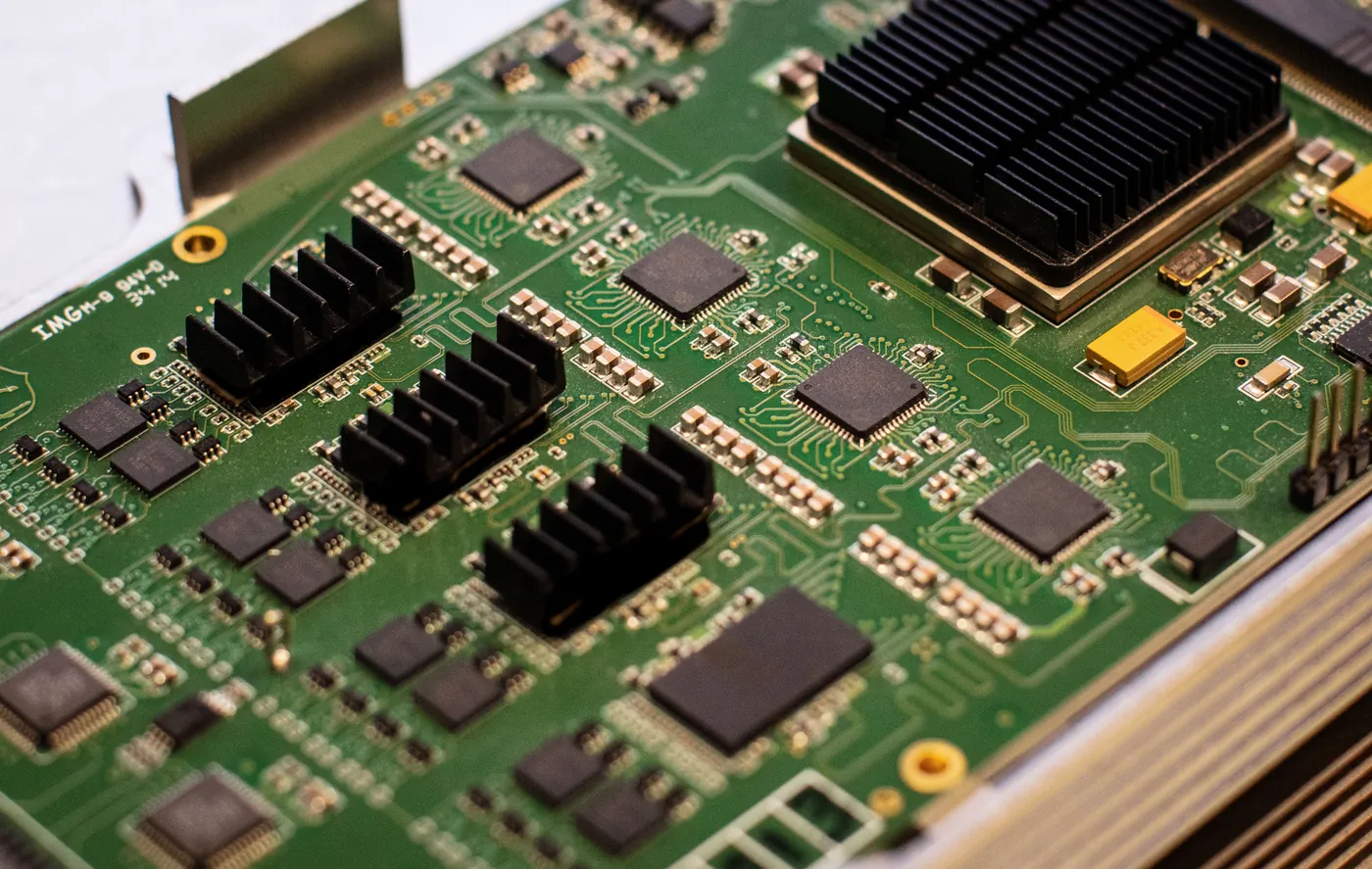
The power of the UltraVision comes from three processors in ascending order of speed: an Intel i7, a graphics processing unit and a very large field-programmable gate array with several billion transistors.
Engineers use the FPGA architecture to create wide data paths for pipelining and parallel processing, giving up to eight simultaneous beamformers. Faster frame rates come with lower transmit rates, which reduces the patient's exposure to ultrasound energy. Because every data path passes through that one FPGA, upgrades and new functionality are delivered with software alone.
The GPU, programmed in DirectX, creates the high-resolution images in real time. The Windows PC runs the touch interface and brings the internet with it: teleradiology, secure patient information handling and the ability to reprogram and upgrade every device remotely.
See the UltraVision XS in action
Online demonstrations are available, with one-time purchase and lease options.