
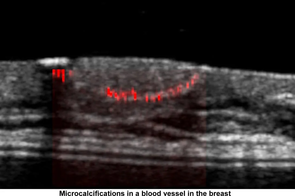
Why calcifications are hard to see
Acoustic impedance is a physical property of the tissue the sound passes through, a measure of how much resistance an ultrasound beam encounters. Tissue has an acoustic impedance of around 1.6 to 1.7 MRayl; a calcium particle is 8 to 20 MRayl. The reflections that make up a B-mode image, between one tissue and another, usually return less than 1% of the transmitted energy, while the reflection from a calcification can return up to 80%.
Calcifications therefore appear as strong white dots. But to produce a cosmetic grayscale image the strong echoes are compressed, and the calcifications disappear into any echogenic volume of tissue. Many researchers have tried to improve the imaging of microcalcifications, and the smaller the particle the more easily it is lost in tissue backscatter.
A different idea: watch them move
Our motivation was to visualize the microcalcifications in the breast that mammography relies on. To be diagnostically significant they are under half a millimetre across, which is why they are called microcalcifications (MCs).
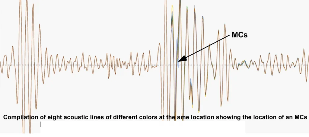
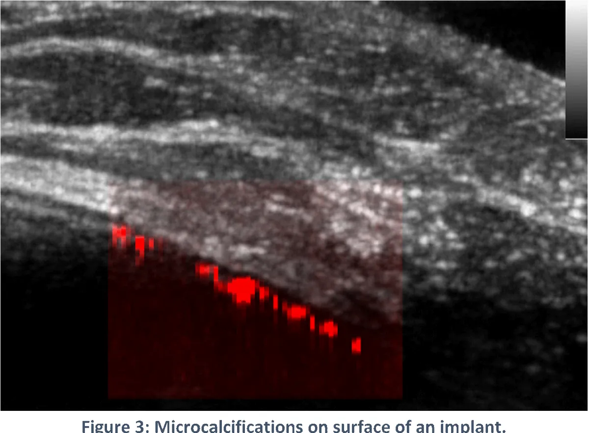
Instead of asking how strongly a particle reflects, MC-mode asks how much energy it absorbs. A microcalcification struck by an ultrasound pulse is pushed a few microns and springs back, while the tissue around it barely moves at all. By sending the same pulse again and comparing the returning echoes, MC-mode keeps only what changed between the two, and in that brief interval the only things that moved were the microcalcifications. Everything else, including blood flow and breathing, falls away.
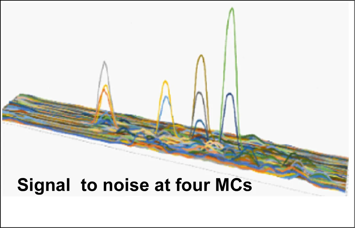
The result is a signal that does not depend on the particle's echo standing out from the tissue around it. That is why MC-mode finds particles that are invisible in B-mode, and finds them with a very high signal-to-noise ratio. The mode uses ordinary diagnostic frequencies and does not change the system's acoustic output.
Results so far
The method has been tested in phantoms with clusters of 50 to 200 micron Type II MCs mixed with scatterers, where it returns strong signals only from the MCs. It has found every MC implanted in chicken and pork breast, and in phantoms it has detected MCs down to 45 microns. So far the method has produced no false positives and has detected every known MC, whether in a breast or placed in a phantom.
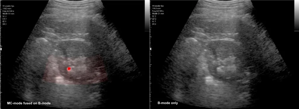
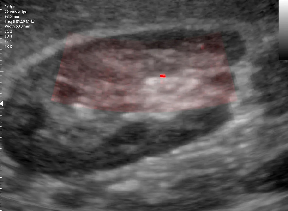
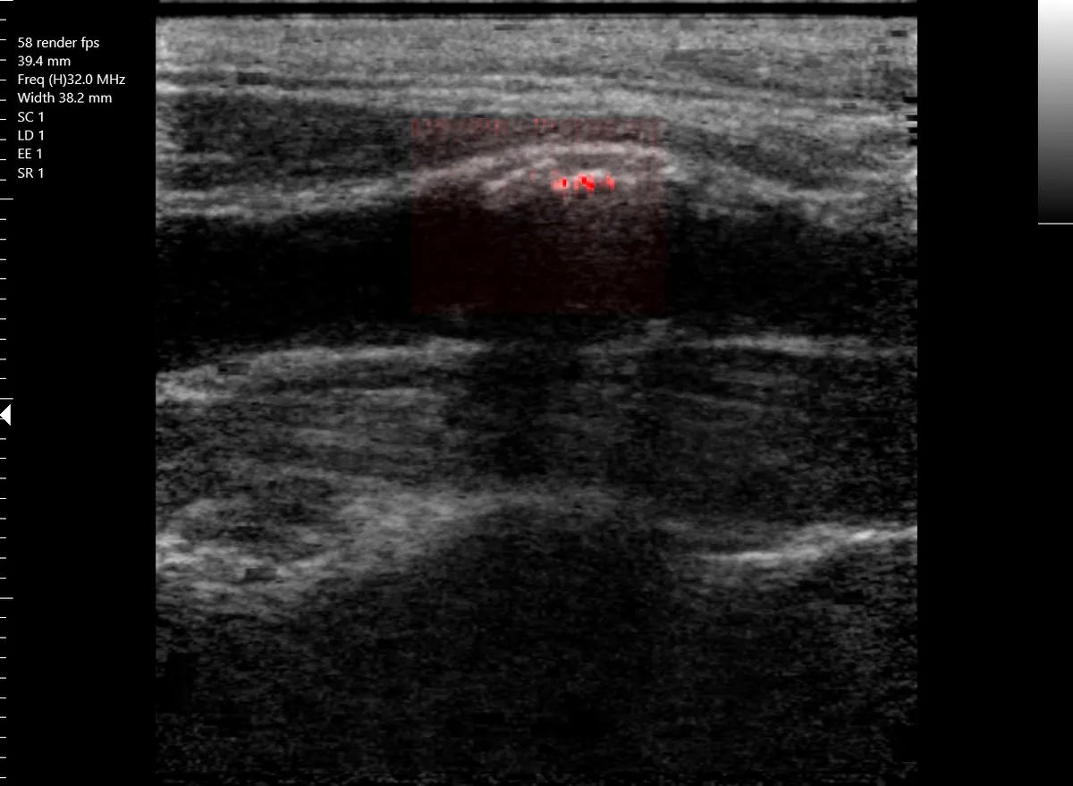
Unexpectedly, the same mode shows kidney stones. In an older patient scanned from the back with a curved array, a stone more than 2 mm across was clearly visible at 75 mm depth: a larger stone subtends more acoustic lines and also moves, so the high signal-to-noise compensates for size. In a younger member of our own team it picked out a tiny stone. It has also highlighted calcified plaque in a carotid artery.
Breast cancer and the two kinds of calcification
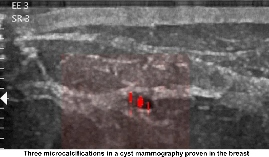
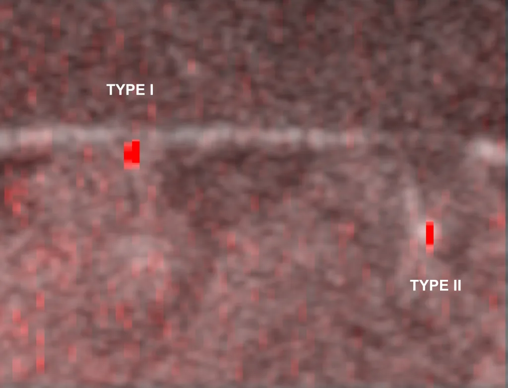
Microcalcifications detected by mammography are responsible for finding most cases of ductal carcinoma in situ, an early form of cancer, and the morphology and distribution of the MCs as defined in the BI-RADS atlas indicate the risk of malignancy. MCs associated with cancer are small rounded crystals of calcium hydroxyapatite (Type II) under 500 microns that appear as white flecks on a mammogram. Calcium oxalate MCs (Type I) also occur in the breast and appear to be a reliable sign of a benign lesion. Mammography cannot distinguish the two, and in B-mode ultrasound both are easily obscured in hyperechoic breast tissue.
In the phantom image above both particles are visible, and MC-mode can be tuned to show either one on its own. The next goal of our research is to tell the two types apart reliably, from 40 to 200 microns in diameter, so that ultrasound can indicate not only where a calcification is but whether it is likely to matter.
Join the MC-mode research. Anyone with an interest in participating in this work, clinically, commercially or as an investor, is urged to contact us.